How do We Sense The evolution of simple cell organisms to
more complex ones resulted in the arrival of humans on Earth. We know that all
organisms have the ability to perceive the environment for survival. Anybody who can perceive or feel things is
sentient. Knowledge of life's physical makeup can help us to determine what
makes us sentient. All sentients perceive external stimuli through sense
organs. Humans have five sense organs: eyes, ears, nose, mouth, and skin. Each sense
organ converts any stimulus into a signal and conveys it to the brain. The brain
analyzes the received signal for sensory data. Through a process called
perception, we get aware of something we sense. The brain also interprets the received
information to make sense of it. The process of thinking and using knowledge is
known as cognition. We will describe the sensing mechanism to explain its role
in a thought process leading to perception and cognition. The Sensory Organs The sensory organs send signals to the
brain as input and receive back signals from the brain as output. The brain-generated
signals can be either to the sensory organs or other body parts. Some examples
of these signals are as follows. The eyes act for the sense of sight and
provide input to the brain for whatever we see. The eyes process the brain
output through movements of various eye parts in reaction to the inputs and
internal thoughts. Similarly, the mouth and tongue are used for eating while
providing a sense of taste as input to the brain. As an output of the brain, the
mouth and tongue perform the movements of the muscles during eating. The
nostrils convey the sense of smell to the brain as an input. While as an
output, nostrils accept the brain signals for sneezing and other nasal
expressions. Similarly, the ears provide the sense of hearing as an input
signal to the brain. The skin gives us a sense of touch, texture,
pressure, and temperature as inputs to the brain. An example of the brain
output to the skin is raising hairs in case of threatening conditions or sudden
pressure or temperature changes. Another significant output of the brain is
moving the muscles to coordinate various activities of different body parts. For
example, the mouth and throat process the brain's output for the act of speech
in response to internal thought signals. Also, just thinking of some foods can
activate salivation, a brain output processed by the mouth in reaction to
internal thought signals. Some activities, such as breathing and
heart-beating, happen all the time and are controlled autonomously by the
brain. As we mentioned earlier, our body parts
are made from cells. Each sensing organ has many sensing cells to convert
stimuli into a neural response. These sensing cells are also known as receptor
cells. For each type of sensing stimulus, the receptors are different. For
example, eyes have photoreceptors that detect light. Ears have mechanoreceptors
for detecting the pressure of sound waves. The nose has chemoreceptors for
sensing different smells. Similarly, the tongue has chemoreceptors
for detecting different tastes. The skin has many types of receptors. For
example, it has mechanoreceptors for feeling pressure and touch. Also, it has
thermo-receptors for detecting hot and cold. In addition, it has nociceptors
for the pain. The receptor cells are coupled to the
nerves to carry the signal to the brain. The brain cells are specialized cells
known as neurons. The nerves connect the body parts to the neurons in the brain.
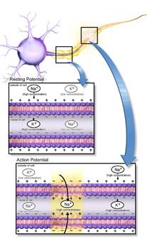
Now, First, we will describe the working principle of the neurons. A neuron cell is shown in Figure 3.1. A neuron cell body encloses the nucleus and other
organelles like other cells. However, it has many spiny structures, as shown in
the figure. The body and spines of a cell are demarcated by the cell membrane. A
cross-section of the cell membrane is shown in more detail in the middle of the
figure. The cell membrane is made of lipid molecules. Outside the cell
membrane, the liquid mixture around the cell has a higher concentration of sodium
ions. On the other hand, the solution inside the
cell, cytoplasm, has a low sodium ion concentration. However, potassium ion
concentrations in the liquids outside and inside the cell membrane are the opposite
of sodium concentration. As a result, we have a potential difference across the
cell membrane. Typically, it is about -70 millivolts. Due to the potential
difference, the cells are called polarized. The cell membrane is excited when a
stimulus above a threshold value is applied to any membrane patch. The membrane
potential is suddenly reversed to a high value by a flow of sodium ions into
the cell, as shown at the bottom of the figure. The cell is depolarized as the
potential difference across the cell wall is reversed. The potential difference
or voltage rises to +40milliVolt. However, the potassium ions inside the
membrane counteract to balance this inflow. In fact, these overreact, causing
repolarization even below the resting potential. However, after a refractory
period, the cell reaches its resting state. Figure 3.1 A
cross-section of the cell membrane of a neuron cell of the nervous system. Credit: Blausen.com staff (2014). "Medical gallery of
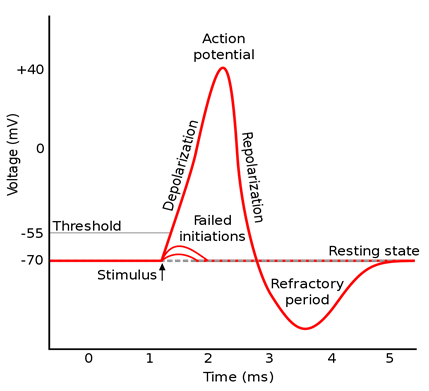
Blausen Medical 2014," [https://creativecommons.org/licenses/by/4.0/] A plot of the change in the membrane
potential difference with time is shown in Figure 3.2.
The shape of the curve, as
shown in the figure, is a typical pulse of the action potential. The action
potential is generated when a stimulus is above a threshold value, as shown in the figure.
The neuron uses these pulses to transfer information. Figure 3.2: A typical
plot of the action potential generated by receptor cells. Credit: [CC BY-SA 3.0 (https://creativecommons.org/licenses/by-sa/3.0)]. The receptor cells are similar to the
neuron cells. The primary function of receptors of the sense organs is to
convert any stimuli into electrical neural signals. An electrical pulse is generated
in response to a stimulus applied to a receptor cell of a sensory organ of the
body as an action potential. Action potential pulses are produced as long as
the applied stimulus to any sensory organ is held above a threshold value.
Thus, a signal consisting of a train of action potential pulses is generated. All signals to and from the brain are in
the form of a train of action potential pulses. For moving any body organ, the
brain keeps sending action potential pulses until the desired position of the
part is reached. These signals pass through the nerves to various parts of the
body. Thus, the nerves carry the input and output signals as electrical pulses.
The mechanism for pulse signal transmission to the brain is the same in all the
sentients. The brain is the central controller of all
the activities of a human or an animal. It is connected to every body part through a network of nerves.
The nerves convey information to the brain and carry back the commands from the
brain. First, we will mention
the critical parts of the brain to understand the processing done by the brain. A diagram illustrating the major parts of the
human brain is shown in Figure 3.3. Four major parts of the brain are the cerebrum, the
cerebellum, the diencephalon, and the brain stem. The cortex is the outer layer
of the cerebrum. The cortex is the main organ responsible for cognitive
processing. Most of the thinking process happens in this part. Thalamus is the uppermost part of the
diencephalon. The thalamus acts as a relay center for most of the sensory
information. Under the thalamus, the hypothalamus is another crucial part of
the diencephalon. The hypothalamus serves as a control center for many of the
autonomic functions. The cerebellum at the back of the brain is
responsible for coordination and balance. In addition, the brain stem is the
control center for essential functions such as breathing and sleeping.
Midbrain, pons, and medulla oblongata are the significant components of the
brain stem.
Credit: Blausen.com staff (2014). "Medical gallery of
Blausen Medical 2014," [https://creativecommons.org/licenses/by/4.0/]. The nerves carry signals from all of the
sensory organs to the brain. The thalamus, a part of the brain, receives these
signals. An exception to this is the smell signal that goes directly to the
primitive cortex of the brain. Other parts of the brain analyze these signals
to extract the information. Based on the analysis, the brain issues command signals
for the nerves to perform various activities. In every part of the brain, the blood is
supplied by the arteries. For the functioning of the brain, all the neuron
cells require oxygen to keep themselves alive. The oxygen is provided by the
blood circulating in the brain arteries. The oxygen-carrying blood to the brain
is pumped by the heart. We will briefly explain the heart's function as it is
essential for the brain's functionality. It is well known that the heart is
situated between the lungs in the torso of a human body, while the brain is
located inside the skull in the head. The heart is a pump supplying blood to
every body part, including the brain. Almost one-fifth of the total blood
pumped by the heart goes to the brain, while the rest is pumped to the
remaining parts of the body. The brain and heart are connected strongly
by the blood supply. The brain needs oxygen continuously to carry out a process
of thinking which is supplied by the blood pumped by the heart. The oxygen
demand varies depending on the requirements of a thinking process. The heart
continuously meets the brain's oxygen demand by changing its pumping rate. The pumping
rate can be observed as the beats of the heart. The heart performs two types of
circulation to supply oxygen-rich blood to the body continuously. Firstly, the
heart performs circulatory functions to provide oxygen-rich blood to all body
parts except the lungs through arteries. The veins bring back the
oxygen-deprived blood to the heart. This circulation of blood is known as
systemic circulation. Secondly, the heart sends oxygen-deprived blood to the
lungs and gets back oxygen-rich blood. This circulation of blood is known as
pulmonary circulation. The heart performs these two types of circulation
simultaneously, as explained below. As shown in Figure 3.4,
the heart has four chambers: two atria and two ventricles. The oxygen-deprived
blood from the body is collected by the veins and is brought into the right
atrium. The collected blood from the right atrium flows into the right
ventricle, which sends it to the lungs to get oxygen. The oxygen-rich blood
from the lungs is returned to the left atrium. From the left atrium, the
purified blood goes to the left ventricle. From there, it is pushed into the
main artery, the aorta, for circulation in the body. In this manner, the heart
can perform systemic and pulmonary circulation simultaneously and continuously,
which is made possible using the four chambers. Also, a simple, alternate
contraction of the atria and ventricles results in the blood pumping in the
order as explained. As explained below, the pump is driven by electric pulses
of the action potential. Figure 3.5: An
illustration depicting the various parts of a heart. Credit: HTTPS://cnx.org/contents/FPtK1zmh@6.27:
MCgS6S0t@3/Cardiac-Muscle-and-Electrical-Activity [https://creativecommons.org/licenses/by/4.0/] For the electrical activity of the heart,
several small nerves are spread throughout the heart muscles. The major
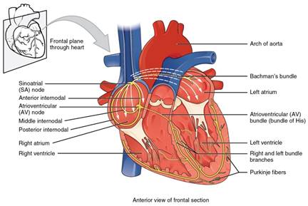
junctions of the nerves are known as nodes. As shown in Figure 3.5,
the sinoatrial node in the heart is the starting point for the depolarization
wave caused by an action potential. Then, the wave reaching the
atrioventricular node spreads to the internodal pathways. Thus, an action potential pulse originating in the sinoatrial node
in the heart's right atrium starts the depolarization resulting in the
contraction of both atria. After
that, the wave goes to the atrioventricular bundle. Then, the wave spreads to the
bundle branches on the left and right. Finally, it reaches the Purkinje fiber. In this manner, the initial wave spreads and reaches the
ventricles resulting in their contraction. The heart and the brain The heart and brain communicate through
four different interactions, thereby influencing the function of each other
continuously. The primary communication between the heart and brain is through
the transmission of nerve impulses of the action potential. Also, the heart and
brain communicate biochemically via hormones and neurotransmitters. In
addition, both interact through pressure waves and electromagnetic fields. In various experiments, it is observed
that an isolated heart continues pumping action as long as it gets oxygen. This
fact is used to keep the heart alive while performing surgery for heart
transplantation. The sinoatrial node can generate action potential pulses
independent of the brain or other body components. However, the brain continuously
regulates the heartbeat rate through a feedback loop. Depending on the
requirement of the given situation, the brain can slow down or accelerate the
processing of current thought. The processing rate, in turn, requires a change
in oxygen demand. The brain slows down or ramps up the heart rate accordingly. The brain controls the emotional response
in addition to rational thinking and analysis. The brain preserves the outcomes
of the thinking process in memory that becomes part of our belief system. An
emotional response is invoked if there is an unexpected sudden change in
something against our belief system. The emotional response is often strong
enough to overtake the rational thinking process. For any emotional response,
the heart rate changes depending on the sudden oxygen requirement of the brain.
From the outside, we immediately observe a difference in heart rate, although each
impulsive thought-invoking action happens in the brain. Therefore, in the
literature, several activities are attributed to the heart instead of the brain.
We will illustrate this further with an example. Generally, our everyday activities are driven
by desires and the need for survival. For example, the need to procreate gives
rise to the urge for love. Love and romance are also brain activities, although
we feel these as a change in the heart rate. Romance is an emotional response.
Most of the existing literature describes the emotional action of romance,
attributing it to the heart. However, the thought process about romance happens
in the brain. For the act of romance, no distinction is made between the heart
and brain in the literature. However, the functioning of the heart and brain are
distinct, as we understand from modern science.Neurons
The brain
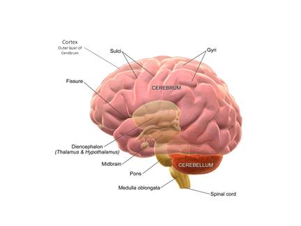
Figure 3.3: Major parts of the human brain.
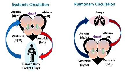
Figure 3.4: A
simplified schematic diagram depicting the heart's blood circulation.